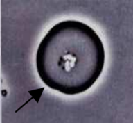
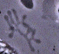
Live Cell Microscopy - Examples
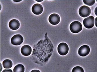
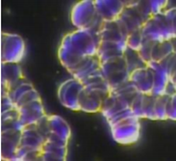
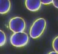
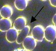
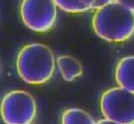
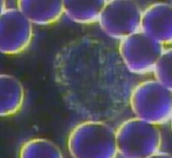
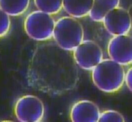
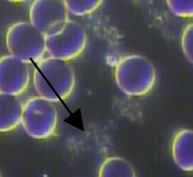
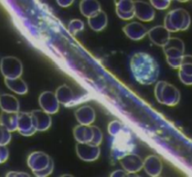
Example of good, healthy red blood cells and environment (following 2 x images)
|
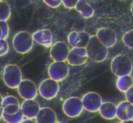
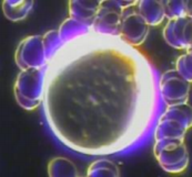
The Red Blood Cells (Erythrocytes) are uniform in size and shape and appear as round circles on a gray background (in Phase Contrast). The center of each cell is lightened somewhat and slightly off white in color. They reside freely in their own space, not overlapping or sticking together, but gently bouncing off each other.
|
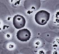
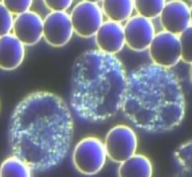
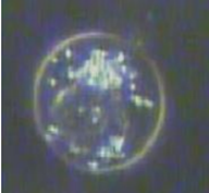
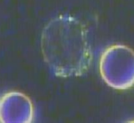
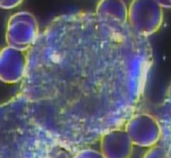
The White Blood Cells are about as large as 2 Red Blood Cells and have a rather grainy
appearance with different characteristics depending on their type. They display many different shapes and some are active and moving. In normal blood there are about 700 Red Blood Cells to every White Blood Cell. |
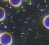
The Plasma surrounding the cells, is clear with some Chylomicron and Globin particles
dancing about. There is also a scattering of platelets but without much cellular breakdown, bacteria, clots, or other undesired floating masses. |
|
|
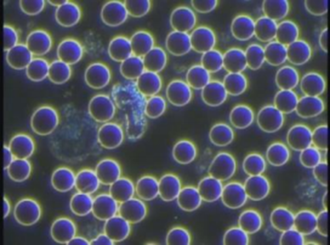
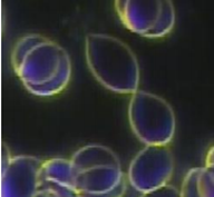
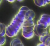
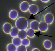
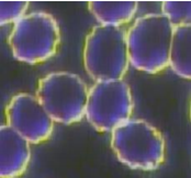
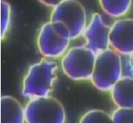
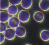
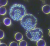
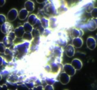
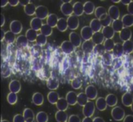
The next 3 pictures are progressively worse indications. They reflect a loss of “electrical charge” of the RBC’s which causes them to stick together. Cells find it more difficult to transfer oxygen to tissues and to circulate freely. CAUSE: Too much cooked animal protein in diet, low digestive capacity, low HCL production and enzyme utilization dehydration, diet not proper for blood type, consumption of processed foods, high sugar and/or carb, prescription/street drugs, excess alcohol intake, smoking, heavy metals. Minerals may be off. Inflammation marker. SYMPTOMS: Fatigue, lethargy, heart stress,poor circulation, BP off, possible edema, blood sugar fluctuations, hormonal imbalances, etc...
Protein Linkage
|
Rouleau
|
Aggregation
|
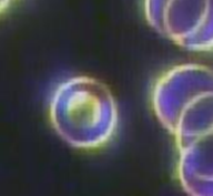
Anisocytosis Variation in RBC size
Macrocytes - Big RBC’s
CAUSE: Overall lack of good nutrition or assimilation which can lead to being low in Vitamin B12, Folic Acid, fat soluble Vitamins A & E. Can be from poor digestion & possible lack of HCL stomach acid, low bile flow from liver dysfunction, low enzyme production. This can lead to lack of friendly intestinal bacteria. SYMPTOMS: Inability of RBC’s to adequately carry oxygen – shortness of breath, fatigue, light headedness, various types of anemia.
Microcytes - Small RBC’s CAUSE: Low in Iron, B12, Folic Acid, same as above. SYMPTOMS: Anemia, low energy, tired. Bone problems, osteoporosis.
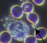
Various Types of Poikilocytes (Variation in RBC shape)
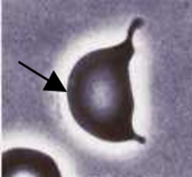
Dacryocytes “Tear shape”
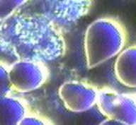
Cells appear in the shape of a teardrop or pear with a single short or long protrusion. If not artefact, some of the tear drops will point in different directions. CAUSE: Often seen in bone marrow disorders but may also be seen in severe forms of anaemia. May indicate low digestive capacity & enzyme utilization. Consider blood type compatibility with regards to diet.
Echinocyte “Spiky shape
RBC’s with sharp burrs/spikes with regularity. They appear shrunken and dying. Up to 1% is normal. CAUSE: Free Radical damage caused by: prescription or street drugs, toxins (environmental or in food), alcohol, nutritional deficiencies from malnutrition or poor diet. Spleen and liver stress. SYMPTOMS: Poor circulation, low energy, degenerative disease conditions.
Ovalocyte “Oval shape”
CAUSE: Lack of Vitamin B12, Folic Acid
(B9) and other B Vitamins. Hormonal imbalance with the Glandular/Endocrine system. SYMPTOMS: Depression, fatigue, dizziness, PMS or other hormonal issues, anemia. |
Burr Cell “Bottle cap shape”
RBC’s with corrugated membrane. CAUSE:Low EFA’s (good fats) in diet, poor digestion too many “bad fats”. Free Radical stress from: diet too low in antioxidants, food additives, sodas, nitrates, fried foods, environmental toxins, food toxins, air fresheners, cleaning products, cigarettes, pesticides, chemicals, metals, street or prescription drugs. SYMPTOMS: Poor circulation, low energy, premature or accelerated ageing.
Acanthoycyte “Irregular spikes”
RBC’s with irregular burrs or spikes. CAUSE: Free Radical damage caused by: prescription or street drugs, toxins (environmental or in food), alcohol, nutritional deficiencies from malnutrition or poor diet. Spleen and liver stress. SYMPTOMS: Poor circulation, low energy, anemia, degenerative conditions.
Codocytes “Target shape”
RBC’s whose membranes have collapsed on itself. CAUSE: Possible low iron or disorganized cellular iron, bile insufficiency, weakness in liver or spleen. SYMPTOMS: Anaemia low B12 and Folic Acid (B9), fatigue, increased cholesterol, passing gas.
|
Elliptocytes “Pencil shape”
Red cells that are elliptical in shape. May range from pencil to cigar shape. CAUSE:Large numbers are most commonly seen in iron deficiency.....possibly poor iron absorption from low HCL production due to stress or strong emotions. They may also be seen in hereditary elliptocytosis.
Various Types of Schistocytes (Damaged or fragmented cell membrane)
Schistocytes “Damaged or fragmented”
Fragments of RBC’s resulting from a
destruction of the cells (broken membrane). CAUSE: May be a sign of the liver detoxifying or under stress. Also seen in cases of oxidative stress due to ingestion or inhalation of toxins, fumes, tobacco, drugs, smog or other free radical promoting elements, exposure to chemicals such as arsenic, lead, enzene, nitrites, and potassium chlorate. SYMPTOMS: Anemia, low energy, tired, degenerative disease. NOTE: May also be seen in severe burns, artificial heart valves, haemolytic anemias, and disseminated intravascular coagulation. Keratocyte “Horn appearance”
CAUSE: Damage to cell membrane occurs when fibrin is being deposited within blood vessels as in disseminated intravascular coagulation -DIC. The circulation is seeing internal coagulation which shouldn’t be.Vascular prosthesis. SYMPTOMS: Circulation issues, cold hands and feet, fatigue, anemia.
|
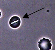
Helmet Cell
Semicircle or crescent shaped RBC’s. CAUSE: Due to low bile salts and low unsaturated fatty acids (EFA’s & UFA’s). Liver and gall ducts may be plugged. SYMPTOMS: Gallstones, indigestion, gas. May also be a sign of a missing or malfunctioning Gall Bladder.
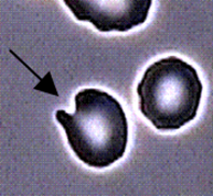
Protrusion
Similar shape as Tear Drop, except with
rounded edge. CAUSE: Due to low bile salts and low unsaturated fatty acids (EFA’s & UFA’s). SYMPTOMS: Gallstones, indigestion, gas. May also be a sign of a missing or malfunctioning Gall Bladder. |
Bite Cell
RBC’s from which denatured, precipitated masses of haemoglobin have been pitted by the spleen. The bites can range from large "bites" to tiny "nibbles". CAUSE: Prescription/street drugs, viral activity or low oxygenation.SYMPTOMS: Chronic fatigue.
Other Red Blood Cell Anomalies
Parasitized RBC “Lesions on RBC’s”
RBC’s with rod formation inside the
membrane. The RBC’s appear “parasitized” (appears as a bacteria inside the cell). They may be infected with rod and tube forms or embryonic bacteria. It can also mean the cells are internally and externally parasitized. They may also have invisible threads in the membrane. If they are not stopped by the immune system, they will continue to attack other cells. CAUSE: May signify low immunity, kidney stress or that the pH is not balanced. The shift in internal terrain creates an environment for microorganisms to invade the RBC’s. May be seen in individuals with autoimmune type diseases and neoplastic disease. Seen in most individuals but is not considered serious unless present in high numbers.SYMPTOMS: Anemia, tired, low energy, fatigue, possible degenerative or auto-immune diseases. Hemolyzed RBC “Ghost Cell”
Round shadowy circles the same size as RBC’s. They are RBC’s that have lost their bilayer and have dispersed haemoglobin leaving a shadow-like appearance. CAUSE: May signify premature red cell destruction from oxidative stress, drugs, alcohol or
smoking. Can also be due to poor nutrient assimilation (especially EFA’s) and dysbiosis. SYMPTOMS: Low energy, depressed, anemia, presence of antibodies, etc... |
Reticulocyte “Premature RBC”
Premature and larger than normal RBC’s with remnants of nucleus. Up to 1% is normal.CAUSE: Blood loss anemia (after haemorrhage), fibroid tumours, iron deficiency, new to high altitudes. SYMPTOMS: Tired.
Heinz Bodies
Inclusions within red blood cells that are
composed of denatured hemoglobin. They appear to have “granules” in the RBC’s - the hemoglobin is damaged. CAUSE: Seen in cells of the circulating blood in people with hemolytic anemias of diverse origins. Also caused by certain prescription drugs. SYMPTOMS: Fatigue, low oxygen. NOTE: May be more easily seen in phase contrast or in labs with a special stain. Flickering RBC
RBC’s appear to “flicker”. Similar to Target cells but not so defined. This appearance is sometimes exhibited in the RBC’s when they are parasitised by micro-organisms They are thinner and deficient in iron. Therefore they have less haemoglobin and cannot carry as much oxygen throughout the body. Often seen along with Target Cells. CAUSE: May indicate low iron, B12/folic acid deficiency, insufficient bile production, anaemia or hypothyroidism. SYMPTOMS: Pale skin, lethargy and tiredness.
|
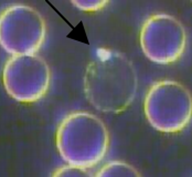
White Blood Cells
Overall WBC count Normal count: There should be approximately 1 WBC for every 700 RBC. Abnormal count: NOTE: This is a perspective based on the
observation of the client’s blood. - WBC’s – FEWER than normal *This is only problematic if there are other issues going on. Make sure to take the “whole”
picture into consideration. Indicates a more catabolic state. WBC’s – MORE than normal CAUSE: Viral activity, bacterial infection, microbial infection, lack of sleep, stress (emotional, physical, environmental). Indicates a more anaoblic state. After eating, there may be an elevated WBC count typically in the following: NONE from raw organic food. MILD from stovetop or oven cooking. MODERATE from pressure cooked or canned food. SEVERE from processed foods, food additives, candy, refined carbs, sugar, soft drinks, etc…WHOPPING from processed meats & microwave cooking.
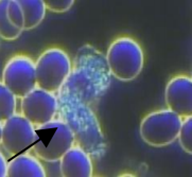
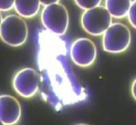
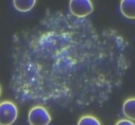
Live blood video showing a white blood cell (neutrophil) chasing bacteria
You will observe this type of activity (on the big screen) when we look at your blood!
Neutrophils
Should make up 65% of the total WBC
count. Neutrophils ingest and destroy bacteria and ther pathogens. Neutrophilia: Increased number of Neutrophils. CAUSE: One basic cause of a higher than normal Neutrophil count is when a high level of stress is placed on the body.Potential causes of high levels of stress include: Nervousness, vigorous exercise, seizures (involuntary muscle movements due to over excitement of nerve cells in the brain), a sudden infection from bacteria, damage or inflammation of tissues, sudden kidney failure, etc…Neutropenia: Decreased Neutrophils. CAUSE: Nutritional deficiencies, Free Radical Damage, Prescription/street drugs, Toxins – from food and environment, Bone marrow damage, auto-immune reactions, radiation and chemotherapy, etc… Neutrophils – Hypersegmented

They have 5 or more nuclei (lobes in the nucleus). CAUSE: Low B-12/folic acid, poor digestion, lack of absorption of vitamins A & E, intestinal dysbiosis, mineral deficiency or imbalance, weakness in liver, kidney or spleen.
Could be congenital and may also be a sign of early bone marrow depletion. SYMPTOMS: Anemia, deficient immune system (reduced ability to destroy parasites or diseased tissue), depression, PMS, etc... Eosinophils
They normally make up 1-4% of the white blood cell count. They are characterized by having large, moving granules which can be highly refractile (able to deflect light) and phagocytic (absorbs wastes and toxins through its membrane). These cells usually contain a bilobate (two lobes) nucleus or the nucleus may have a kidney shape to it. In Darkfield Microscopy, they appear very bright and shiny. NOTE: Eosinophils with white edges indicate an allergic reaction or allergies. Eosinophilia: An excess of Eosinophils indicates an anabolic state. CAUSE: Allergies, edema, eczema, reactions to parasites, drugs (street or pharmaceutical), smoking, second hand smoke, bone bruises, irritation, inflammation, menstruation, arthritis, etc…SYMPTOMS: Irritation, soreness, inflammation, pain, aches, swelling, sinus congestion, drainage, circles under eyes, etc...NOTE: In this state, the Eosinophils have round non motile coarse granules concentrated on the outside. Decreased Eosinophils: Indicates a catabolic state. CAUSE: May be due to administration of steroids or other prescription or street drugs. SYMPTOMS: Irritation, soreness, inflammation, pain, aches, swelling, sinus congestion, drainage.
|
Neutrophils – Inactive
They appear rounded and their granules are not streaming. CAUSE: Altered blood pH, compromised immune system, mineral imbalance, B12/Folic Acid (B9) deficiency, malabsorption, not enough exercise, too much alcohol or sugar, yeast imbalance and other infectious states. This may also happen if the slides are cold. SYMPTOMS: Low Immune system which creates a lower ability to destroy parasites and infected or diseased tissue.
Neutrophils – Band Cells
Known as “young” Neutrophils that have a nucleus in the shape of a horseshoe
(unsegmented nucleus). CAUSE: They appear in larger numbers in the bloodstream in response to stress or infection. Basophils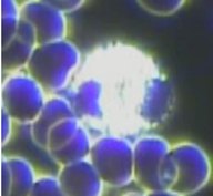
They make up less than 0.5 per cent of the white cell count. They contain large
cytoplasmic granules and are also phagocytic. They can secrete a biologically active substance such as histamine and heparin (chemicals which induce inflammation). In Darkfield Microscopy, they appear to be highly reflective and resemble Eosinophils except they are more round and smaller in shape. In Phase contrast their granules are seen to be larger than those of Eosinophils. Basophils are often associated with asthma and allergies. Related cells, known as mast cells, are often associated with helping provide mediators to initiate immune responses. Basophilia: When there is an excessive amount in the blood (more than 2% of the white cell count), they have slow moving granules, low activity and are usually nonphagocytic appearing. They release histamine and heparin when activated. CAUSE: Allergies to inhalants, mold, dust, pollen, animal dander, chemicals, perfumes, stressed kidneys, diuretics. May need magnesium chloride. SYMPTOMS: Runny nose and eyes, stuffed up sinuses, flu or cold like symptoms, water retention and edema (swelling). Decreased Basophils: CAUSE: Stress, pregnancy, hypersensitivity reaction, corticosteroids, hyperthyroidism. |
AGRANULOCYTES
Monocytes
Comprise 2-8% of the total white cell count. Monocytes are usually identified by their large bilobate nucleus (1 large nucleus with 2 lobes). They appear “dark” in darkfield microscopy. They resemble Lymphocytes except they are a little bigger and generally have more 5
cytoplasm. In the tissue they further mature into macrophages. Increased Monocytes: CAUSE: Infection (viral or parasitic), inflammatory bowel disease, auto-immune or other degenerative diseases (sarcoidosis, neoplasms, monocytic leukemia, lymphoma, multiple myeloma). Decreased Monocytes: CAUSE: Aplastic anemia, lymphocytic leukemia, intake of glucocorticoids. Lymphocytes with Cytoplasm damage (white areas on edge)
CAUSE: May be due to viral activity, low
oxygen or immune deficiency. High numbers indicate possible bone marrow damage. “Parasitised” Lymphocyte (Bubble surrounding lymphocyte)
CAUSE: Imbalance in the biological terrain (internal environment of the body – Ex: pH). See #10 for potential causes.
12) “Filaments” on Lymphocyte membrane (no picture available) CAUSE: Imbalance in the biological terrain (internal environment of the body – Ex: pH). |
Lymphocytes
B and T cells
There are two types of Lymphocytes: B cells and T cells. They are the smallest of the WBC’s. They comprise 35% of the total white blood cell count. They look very similar to Monocytes but they are smaller. They are formed in the bone marrow or lymph tissues and are approximately the same size as RBC’s. It is hard to distinguish between a B & T cell. They are not very reflective in Darkfield Microscopy. Lymphocytes play a large role in defending the body against disease and are responsible for immune responses. Lymphocytosis: Increase in Lymphocytes. CAUSE: Acute viral infections, infectious mononucleosis, hepatitis, tuberculosis, degenerative disease conditions, free radicals and toxins. There is also an excess when the lymph system is fighting and when there are environmental chemicals, pesticides, food additives, smog or electromagnetic fields (EMF’s). SYMPTOMS: Fatigue, low oxygenation. - Decreased Lymphocytes: CAUSE: May indicate immune deficiency, radiation damage, free radical damage, HIV infection (resulting in AIDS), corticosteroids and other medications that suppress the immune system, malnutrition, problems in bones or by x-rays. “Activated” Lymphocytes (larger than normal)
CAUSE: Imbalance in the biological terrain (internal environment of the body – Ex: pH) from things like: Environmental chemicals, pesticides, food additives, smog, bacterial infection (Mycoplasmas, Rod shaped, LFormed), micro-organisms, street/prescription drugs.
|
Plasma Observations
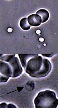
PLATELETS (Thrombocytes)
PLATELETS (Thrombocytes)
Platelets are part of your blood clotting mechanism. When you cut yourself or experience vascular damage, platelets go to the area of the assault and bind together with fibrin so you don’t bleed to death. Platelets can become excessively “sticky” in circulation from some of the following situations. 1) Platelet count NOTE: This is a perspective based on the observation of the client’s blood. Thrombocytosis: When the platelet count is increased. This is seen in conditions such as acute and chronic infections, cancer and certain blood diseases. It may also lead to increased blood clot formation. Thrombocytopenia: When the platelet count is decreased. This may occur as a result of vitamin B12 or Folic Acid (B9) deficiency, liver stress, excessive platelet destruction. NOTE: May also be seen in myelodisplasia or Leukemia. |
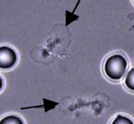
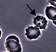
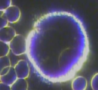
Platelet Aggregation
Platelet clumping or aggregation, which plays an important role in the ratios of
cholesterol/triglycerides/HDL. These excess stickyplatelets contribute to circulatory problems. Large amounts of aggregation can cause poor circulation and narrowing of the blood vessels, which may lead to heart conditions and clots. CAUSE: High fat or triglycerides (especially animal fats) and carbohydrate intake can cause this to happen. May also be caused by excessive red meat, stress, caffeine, sodas, chocolate, and smoking. Often seen in people with diabetes or hypercholesterolemia. SYMPTOMS: Poor circulation, capillary blockage, blood clots, high cholesterol, heart stress, candida and headaches. Foods that help: High sources of Vitamin C (such as Rosehips) help to maintain collagen and strengthen blood vessels. Sources of Vitamin E help to inhibit coagulation. Sources of EFA’s such as fish oils help to improve blood viscosity. |
Pteroharpen “Spreading Platelets”
When platelets are out of the body and put on a slide with a cover slip, the membrane may undergo spontaneous spreading. The area that is spreading is called the Hyaloplasmic Veil. This is a shadowy form, often behind and surrounding the platelets like a haze that make it appear as if the platelets have butterfly wings. CAUSE:This phenomenon is still a mystery but many belief systems associate it with pH imbalance, low oxygenation, leaky gut syndrome (internal toxicity), low friendly intestinal bacteria, antibiotic therapy, high blood sugar, excess dietary PUFA’s or carbohydrates. May also be related to undigested protein in the blood. High amounts of animal protein from cooked animal food sources (or even undigested protein from vegetable sources) in the diet is a common profile. They can signify strong circulatory hindrance. SYMPTOMS: Fatigue.
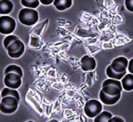
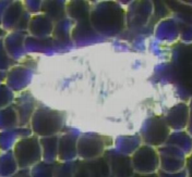
CRYSTAL FORMATIONS
Crystals
Items that resemble glass formations. Some may have colours. They are a sign of high acidity in the blood. They are sclerotic or pseudo crystalline chunks from undigested proteins, fats and acids. They may be seen by themselves or stuck to other blood elements.“Highly Reflective” Crystals. May be a sign of increased cholesterol or high LDL ratios. CAUSE: May be due to eating too many “bad fats” such as deep fried foods. May also be due to liver reacting to high insulin levels from diet too high in simple and processed carbohydrates. If liver and gallbladder are “sluggish” due to too much toxic exposure from food or environment (or toxic emotions), then this may cause poor digestion of fats due to low bile secretion. SYMPTOMS: Increased blood pressure, clogged arteries, plaque buildup.
Coloured crystals
These are an expression of chemicals and metals in the body. Crystals are a combination of various wastes in the plasma that have been there for a while and become hardened over time. They may be from our food, the environment or toxic waste accumulation from poor digestion. CAUSE: May be a sign of
intensified blood fats, undigested protein accumulations, poorly digested foods that sit in the colon, bowel and/or liver toxicity or constipation. May also signify heavy metals in the body. LDL level may be high but it is dangerous if it is low. SYMPTOMS: High blood pressure, clogged arteries, plaque buildup, circulation problems, degenerative disease, auto-immune disorders, arthritis, gout, joint pain (from cartilage damage), bacterial infections, etc…. Crystal Shapes
|
“Broken Glass” Crystals
May be a sign of high Triglyceride ratios.
Signs of atherosclerotic plaque. CAUSE: Same as above. Also may be due to altered pH or low mineral charge. SYMPTOMS: Increased blood pressure, clogged arteries, plaque buildup. Crystal and Toxin Colours
NOTE: The following colours and shapes may appear on their own or stuck to other blood elements.
|
BLOOD PROTEINS
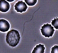
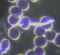
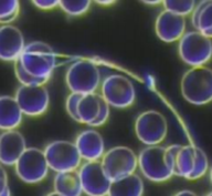
Three blood proteins are albumin, globulin and fibrinogen. A high albumin/globulin or A/G ratio is a sign of good health. We cannot see these proteins in the blood, but we can see fibrinogen when it begins to activate and turn to fibrin. Seeing fibrin strands develop in your picture, depending on speed and intensity, is a sign the blood might be a bit thick and this could be stressful for the liver and cardiovascular system in general.
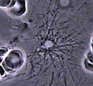
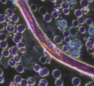
Fibrinogen strands (Filets) “Fibrin Formations”
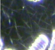
They resemble “cracked” glass. Straight, hairlike formations that look like pick-up sticks in the plasma fluid. The thicker the filet, the more the condition is pathogenic. CAUSE: Can be related to joint problems. Denotes hyperproteinemia, liver stress from toxicity and/or congestion, and associated with toxic bowel, especially when Spicules are also present. Poor digestion and/or bacterial overgrowth can be suspected as cause of bowel toxicity along with old, decaying, impacted fecal matter. Toxins from antibiotics, drugs, alcohol, tobacco, coffee and meat may throw off the pH balance causing liver stress. Seen also in autoimmune diseases such as Lupus, MS, MG, Lou Gehrig's. May also be a degenerative disease indication. SYMPTOMS: Constipation, indigestion, heartburn, bloating, gas, flatulence, fatigue, headaches, backaches, poor circulation, heart stress.
|
Spicules “Dandelion puffs”
Fibrin strands that resemble dandelion puffs. These are altered, unusable polymerized proteins in the blood. They signify liver stress. Spicules could also be a healing indicator if undergoing body cleansing and liver detoxifying. Cause: Toxicity due to alcohol, drugs, antibiotics, tobacco, coffee or excessive amounts of sweets (including excessive fruit consumption). May also signify “heavy proteins” in the blood due to undigested proteins. These toxins throw off the pH balance causing liver stress. Seen also in Autoimmune and degenerative diseases such as lupus, MS, MG, Lou Gehrig's. Foods that help: May benefit from foods high in B Vitamins or Vitamin B-Complex, high enzyme foods such as raw vegetables or Digestive Enzyme supplements, Minerals and Probiotics.
|
Clotting Fibrins
Appears as long strands mixed with thicker blood protein elements. Early signs of clot formations causing thicker blood. CAUSE: Stress, poor protein digestion, incompatible protein for blood type. Liver stress, poor bowel elimination, etc….See Fibrinogen strands for other causes. SYMPTOMS: Poor blood circulation, fatigue, constipation, etc…
OTHER BLOOD ELEMENTS
Plasma Particles “Moving dots”
These are normal blood elements. There should be more if a person has recently eaten and less if a person hasn’t eaten for more than 4 hours. These particles are hard to distinguish from each other without the use of specialized equipment
They may vary from the following elements: Chylomicrons – fat particles • Cell Wall Deficient forms (CWD) – bacterial forms resistant to antibiotic treatments • Protits – protein and lipoprotein particles. Bacterial forms: L-Form, Mesasomes, etc…Cell organelles from cellular breakdown. Mycoplasma Form
A genus of bacteria that lack a cell wall.
Because they lack a cell wall, they are unaffected by some antibiotics such as penicillin or other beta-lactam antibiotics that target cell wall synthesis. CAUSE: Altered pH, stealth infection, stress, lack of sleep, physical strain, overwork, overuse of antibiotics. SYMPTOMS: Fatigue, aches and pains, poor circulation, etc… Myelin Strands
Thin, wispy wormy looking moving filaments. They are polymerizations of blood proteins albumin and globin that form long strings. The organisms multiply locally or spread throughout the body expressing and causing disease. They form on or in the cell and interfere with cellular respiration. CAUSE: Membrane breakdown from all cellular & subcellular particles due to pH imbalance. Generally results from pathologic separation of certain cellular constituents that have high proportion of lipids.
Phospholipid forms “Various stages of Thecits” (as per Enderlein)
Due to acidic pH level which prevents
sufficient oxygen to be transported to the cells, cell death occurs. The lipids (phospholipids) from inside the cell protoplasm are then liberated into the hemoglobin. CAUSE: Dehydration, too much sugar or carbohydrates in diet. Some theories believe that yeast are indicated in advanced stages of latent tissue acidosis. This means that the pH level is acidic which prevents sufficient oxygen to be transported to the cells. This causes cells to die and yeast, bacteria, fungus or mold to grow. Shows over acidity as yeast cannot live in an alkaline environment. May be found in people with cancer, fibromyalgia and those with chronic fatigue. SYMPTOMS: Muddled thinking, slower reactions, sore and flabby (shrinking) muscles, loss of strength, CNS disorders, Alzheimer’s and Parkinson’s. Dark Colour: Correlated to mouth pathology and heavy metals (FOCI – focal area of interference). Light Colour: Appearance of Yeast, fungus or mould in the plasma. Correlated with Yeast and sugar issues. Yeast is a fungus that feeds on undigested food and sugar in the blood. The principal yeast found in the blood is Candida Albicans. This condition is also indicative of over acidity as yeast cannot live in an alkaline environment. |
Bacterial, Rod, LForms or “Parasitic” Forms
Double spore, barbells, rod forms or tiny
worm-like appearance. This is an indication of an imbalanced pH in the body. This bacterial stage indicates immune stressors which if carried to extremes can compromise the health. CAUSE: Immune insufficiency, leaky gut syndrome, various infections in the body, etc... Fungal Forms
Appears as “strands” that sometimes seem to link RBC’s together in the plasma. A change in the terrain (imbalance in the body), creates an
environment for fungal infections to take advantage of a victim’s lowered defenses. Overgrowth of fungal forms shows there is great stress to the body. CAUSE: An imbalance in the body causes the pH to shift and creates a bacterial competition (good and bad bacteria) and allows fungi to multiply. The pH may be shifted due to a weak immune system, treatment with antibiotics or other medication, an infection, lack of exercise, poor diet (or diet not compatible with blood group antigens), negative thoughts, etc....SYMPTOMS: Asthma, allergic alveolitis (inflammation of the lung tissues), diabetes mellitus, various fungal infections, etc.... Myelin Formations
Thicker, expansive, worm like, fungal
appearance. More pathogenic phase than Myelin Strands. CAUSE: Membrane breakdown from all cellular & sub-cellular particles due to pH imbalance. Generally results from pathologic separation of certain cellular constituents that have high proportion of lipids. Uric Acid “Bubbles”
May be seen as “bubbles” or stuck to other blood elements. CAUSE: May be poor protein digestion due to low HCL or Protease enzyme production, eating wrong proteins for blood type or poor digestion from stress or strong emotions. SYMPTOMS: When protein isn’t properly digested, it can turn into uric acid and deposit in joints, causing inflammation. Gout and arthritis are types of joint inflammation.
|
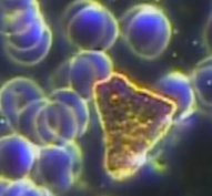
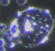
Symplasts
Granular looking chunks with a hard outer shell. Resembles “rock formations” and may appear reflective in Darkfield Microscopy. This is a merge of plasma colloids and particles due to a change in electrical charge once the blood is out of the body and onto the slide. Can cause hardening of the blood vessels and slows down the blood flow. They vary in color and size and are densely packed with toxins and microorganisms. Grey ones tend to show more “protein” issues and white ones tend to show more calcification and plaque issues. When they appear as very dark brown or “black” it is a sign of high toxicity from chemicals in our food and the environment. They may contain the following toxic substances that have adhered to each other: lipid core, fibrin deposits, thrombocyte aggregation, calcification, proteins, lipids, cellular debris and collagen (calcium imbalance). CAUSE:Poor circulation, high cholesterol, mineral imbalance, digestive insufficiencies (low HCL and enzyme production), etc… SYMPTOMS:Pain in calves, cold hands & feet, low energy toxicity, skin disorders, frequent and recurring colds, flu and infection, and possible tooth decay, etc… Commonly seen when the individual is undergoing amalgam removal. It is also commonly seen in someone who is recovering from, or was recently infected by viral forms.
Blue-Grey or Greyish Forms
CAUSE: May be poor protein digestion due to low HCL production, eating wrong proteins for blood type or poor digestion from stress or strong emotions.
Plaque formations “White and reflective
Signs of “Free Calcium” causing high blood calcium. The excess Calcium adheres to other blood elements such as uric acid (from poor protein digestion) and oxidized lipids or cholesterol. CAUSE: May be from supplementing with poorly absorbable forms of Calcium such as Calcium Carbonate. May also be from poor Calcium absorption due to lack of HCL or other elements required for bone formation such as: Minerals, B-Vitamins, Vitamin D. May also be due to endocrine imbalances causing poor thyroid or parathyroid function.
Spheroplast “Round shape”
CAUSE: Similar to Protoplasts.
Thallus “Long and skinny with toxins”
CAUSE: Similar to Capillary Plugs except they are colourful and a sign of high toxicity.
|
Dark Brown or Black Forms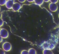
CAUSE: May be a sign of poor protein
digestion over a long period of time leading to a very toxic state. Protoplasts “Jelly-fish appearance”
CAUSE: May be caused by accumulation of toxins from environment, food or undigested food particles. Usually a sign of high bacterial, fungal or viral activity or may also be a sign of parasite (vary in size from large to microscopic).
Capillary Plug “Long and skinny shape”
CAUSE: Toxic build up leading to crystallization in capillaries. The capillaries get weak and burst, releasing these capillary shaped crystals into the bloodstream.
Drepanids
Loose particles that adhere to each other in long skinny rows. They vary in colour from grey, to a blue-grey to white. CAUSE: Can be from poor protein digestion or Calcium absorption from lack of HCL production due to stress or strong emotions. If they are white, it may also be a sign of high acidity forcing Calcium to be leached from the bones and tissues as a pH buffering mechanism.
|
Symplast Shapes
NOTE: The following shapes may appear on their own or stuck to other blood elements. Square: Neurological problems and immune disorders. SYMPTOMS: Nervousness, highly strung, irritability, hyperactivity, depression, listlessness, aimlessness, headaches and migraines.• Trapezoidal: Caused by poor digestion of fats. Circular: Immune system inactive and/or deficient, poor elimination of toxins.